By late January, 1.4 million people in Liberia and Sierra Leone could be infected with the Ebola virus. That’s the worst-case scenario of the Ebola epidemic in West Africa recently offered by scientists at the US Centers for Disease Control and Prevention. The CDC warns that those countries could now have 21,000 cases of the virus, which kills 70 percent of people infected.
One of the big problems hindering containment of Ebola is the cost and difficulty of diagnosing the disease when a patient is first seen. Conventional fluorescent label-based virus detection methods require expensive lab equipment, significant sample preparation, transport and processing times, and extensive training to use. One potential solution may come from researchers at the College of Engineering and the School of Medicine, who have spent the past five years advancing a rapid, label-free, chip-scale photonic device that can provide affordable, simple, and accurate on-site detection. The device could be used to diagnose Ebola and other hemorrhagic fever diseases in resource-limited countries.
 |
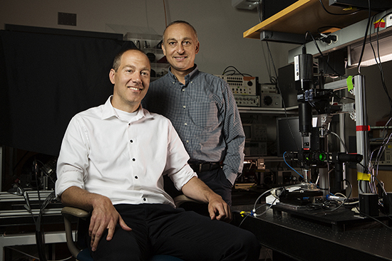
John Connor, a MED associate professor of microbiology (left), and Selim Ünlü, a College of Engineering professor and associate dean for research and graduate programs, have developed a rapid chip-scale photonic device that can detect viruses, including Ebola, on site. (Photo Credit: Steve Prue)
|
The first demonstration of the concept, described in the American Chemical Society journal Nano Letters in 2010 and developed by an ENG research group led by Selim Ünlü, a professor of biomedical engineering, electrical and computer engineering, and materials science and engineering, in collaboration with Bennett Goldberg, a College of Arts & Sciences professor of physics, showed the ability to pinpoint and size single H1N1 virus particles. Now, after four years of refining the instrumentation with the collaboration of John Connor, a School of Medicine associate professor of microbiology, and other hemorrhagic fever disease researchers at the University of Texas Medical Branch, the team has demonstrated the simultaneous detection of multiple viruses in blood serum samples—including viruses genetically modified to mimic the behavior of Ebola and the Marburg virus.
Mentioned in Forbes magazine as a potentially game-changing technology for the containment of Ebola, the device identifies individual viruses based on size variations resulting from distinct genome lengths and other factors. Funded by theNational Institutes of Health, the research appears in the May 2014 ACS Nano.
“Others have developed different label-free systems, but none have been nearly as successful in detecting nanoscale viral particles in complex media,” says Ünlü, who is also ENG associate dean for research and graduate programs, referring to typical biological samples that may have a mix of viruses, bacteria, and proteins. “Leveraging expertise in optical biosensors and hemorrhagic fever diseases, our collaborative research effort has produced a highly sensitive device with the potential to perform rapid diagnostics in clinical settings.”
 |
|
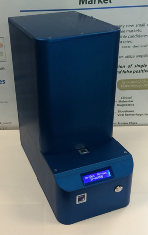
NexGen Arrays prototype of SP-IRIS, which detects pathogens by shining light from multicolor LED sources on viral nanoparticles bound to the sensor surface by a coating of virus-specific antibodies. (Image courtesy of NexGen Arrays) |
Whereas conventional methods can require up to an hour for sample preparation and two hours or more for processing, the current BU prototype requires little to no sample preparation time and delivers answers in about an hour.
“By minimizing sample preparation and handling, our system can reduce potential exposure to health care workers,” says Connor, a researcher at the University’sNational Emerging Infectious Diseases Laboratories (NEIDL). “And by looking for multiple viruses at the same time, patients can be diagnosed much more effectively.”
The shoebox-sized battery-operated prototype diagnostic device, known as the single particle interferometric reflectance imaging sensor (SP-IRIS), detects pathogens by shining light from multicolor LED sources on viral nanoparticles bound to the sensor surface by a coating of virus-specific antibodies. Interference of light reflected from the surface is modified by the presence of the particles, producing a distinct signal that reveals the size and shape of each particle. The sensor surface is very large and can capture the telltale responses of up to a million nanoparticles.
In collaboration with BD Technologies and NexGen Arrays, a start-up based at the Photonics Center and run by longtime SP-IRIS developers David Freedman (ENG’10) and postdoctoral fellow George Daaboul (ENG’13), the research team is now working on making SP-IRIS more robust, field-ready, and fast—ideally delivering answers within 30 minutes—through further technology development and preclinical trials.
SP-IRIS devices are now being tested in several labs, including a Biosafety Level-4 (BSL-4) lab at the University of Texas Medical Branch that’s equipped to work with hemorrhagic viruses. Other tests will be conducted at BU’s NEIDL once the facility is approved for BSL-4 research. Based on the team’s current rate of progress, a field-ready instrument could be ready to enter the medical marketplace in five years.
Find out more about SP-IRIS in this National Academy of Engineering radio clip.
Mark Dwortzan can be reached at dwortzan@bu.edu.


 CN
TW
EN


